The Academy section is a pivotal knowledge center for the farm animal industry, particularly in artificial insemination and veterinary imaging, providing up-to-date educational resources and training. IMV Technologies Academy offers flexible learning options, including technical articles, blogs, on-demand courses, and replay webinars, enabling clients to access the latest advancements in the industry. Expert-led materials across various species ensure comprehensive knowledge enhancement, helping clients stay competitive and improve their technical skills in their respective specialties. This resource is crucial for staying informed and excelling in animal reproduction practices.
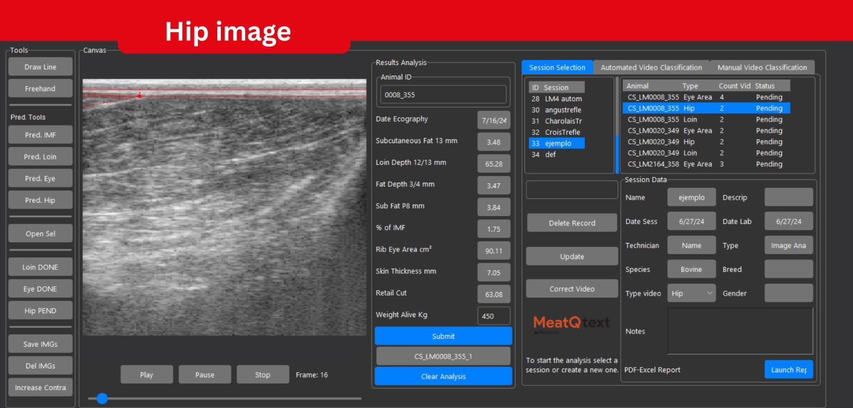
Discover how MeatQtext, the dedicated software for the ExaPad Mini ultrasound scanner, delivers real-time, AI-based intramuscular fat (IMF) measuremen...
Read more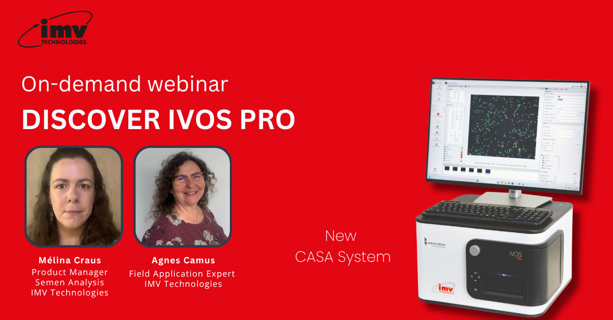
Watch our international webinar and learn how IVOS PRO, our new CASA system, brings automation, precision, and speed to semen analysis. Explore how it...
Read more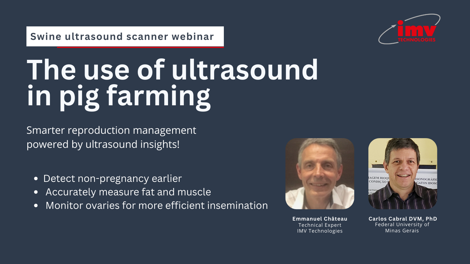
Discover valuable insights from our international webinar on ultrasound in sow reproduction management.In this replay, Prof. Carlos Cabral (Federal Un...
Read more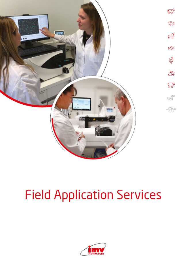
IMV Technologies offers biological training courses, ready-to-use standard protocols...
Read more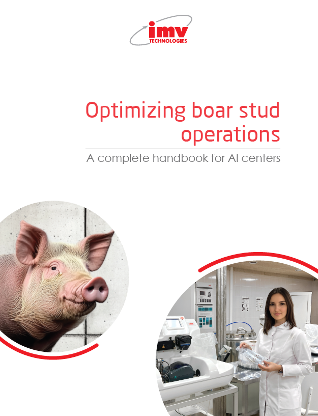
This publication outlines IMV Technologies recommendations for artificial insemination centers....
Read more
Scope:The objective of the training is to give a deeper understanding of milt...
Scope:The objective of the training is to give a deeper understanding of milt...

2 daysREF 026348
2 daysREF 026346